Ocular oncology
Dr Warrier specialises in the diagnosis and treatment of adult ocular tumours. The word tumour essentially refers to lumps either on or within the eye. Patients referred with these lumps, will be assessed thoroughly to exclude the most sinister conditions, which includes melanoma. The latest ocular imaging techniques such as EDI-OCT (enhanced depth imaging – optical coherence tomography), ultrasonography and flourescein angiography may be used to help differentiate malignant from benign lesions.
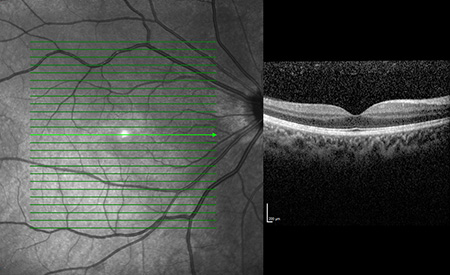
Normal OCT of the macula
Assessment of Ocular Tumours
Aside from his extensive clinical experience in assessing these lesions, Dr Warrier uses the latest technology to differentiate benign from malignant tumours.
EDI-OCT (enhanced depth imaging – optical coherence tomography)
This camera uses lasers and light to show a slice of the retina and choroid. It is very useful for thin lesions in the eye and to detect whether or not they secrete fluid.
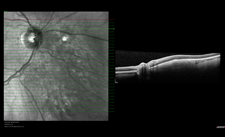
EDI-OCT showing the optic nerve (small bump) and a choroidal tumour lifting up the retina (larger bump).
Ultrasonography
This technique uses high frequency, inaudible sound waves emitted into the eye. These bounce back off any tissue surface towards the probe which measures the ‘loudness’ of the reflected sound and the time taken for the sound to travel into the eye and back again. The intensity of the signal represents the hardness of the lesion and the time taken gives an indication of the distance travelled by the sound. The consistency of the tumour is also assessed.
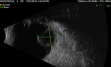
B-scan ultrasound of a choroidal melanoma – It protrudes into the eye and is acoustically hollow.
Flourescein Angiography
This is performed by injecting a flourescent dye into a vein in your arm and taking a series of photos of the back of your eye. This test provides information about the blood supply to the tumour and to the back of your eye. The dye tends to cause a wave of nausea in 10% of patients. Yellow discolouration of the skin and urine occurs until flushed out of the next 48 hours. Approximately 1/2000 people may suffer an allergic reaction to the dye which is very, very rarely fatal (1/200,000).
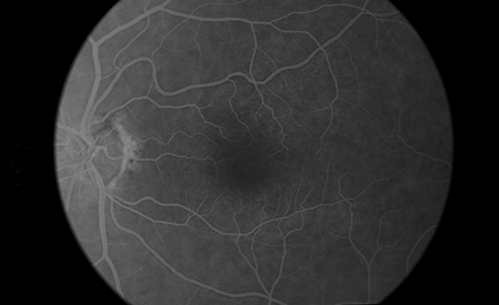
Flourescein angiogram with white dye highlighting retinal blood vessels.